
Ureteroscopy
Ureteroscopy is a common urological procedure administered in patients having urinary tract and bladder related disorders. Ureteroscopy is recommended for patients having kidney stones. The movement of the renal stones is monitored by urologists. Ureteroscopy is a minimal invasive endoscopic procedure predominantly involving the urethra, upper urinary tract and the urinary bladder.
Ureteroscopy Procedure
A flexible or rigid form of telescope is passed through the urethra in order to view the affected region. The procedure is performed under general or spinal anesthesia. The telescopic examination of the ureter and associated organs reveals the exact location of the stone and also other disorders of the system. Other diagnostic tests such as X-rays, CT scan, EKG along with laboratory parameters like urinalysis and complete blood count also help in the effective determination of the underlying condition. This technique is usually followed by the other associated procedures such as lithotripsy in which laser beams are administered to the affected region to dissolve the calculi(stone) or to clear urinary tract strictures. The entire procedure may last from 30 minutes to three hours and patients are advised to stay in the hospital for a day.
The urologist uses the uterescope to remove small kidney stones while larger stones need to be broken up before removal. A kidney stone that has escaped from the kidney and got stuck in the ureter can be pushed back into the kidney. Here it has to be broken into smaller pieces to aid removal. A stent is usually left in place to allow the kidneys to pass the urine to the bladder. This is kept for a few days in case there is swelling and subsequent difficulty in draining the kidney of the urine.
Complications and risk factors
Hematuria happens because of the insertion of the ureteroscope. It usually subsides within 3 days. Antimicrobial therapy is given if hematuria is followed by an infection. Other possible complications are:
Stent associated pain
Perforations caused because of stents
Abdominal pain
Lower back pain
Urethral stricture or perforation
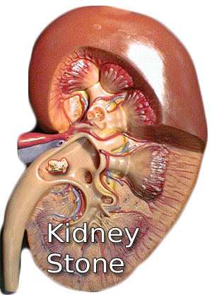
Kidney Stone
A kidney stone or renal calculus is a crystal concentration formed in the kidneys. Nephrolithiasis is formed from the minerals consumed in the diet and is largely composed of calcium. 75% of kidney stones are calcium stones. While Struvite stones are more commonly noticed in women, Uric acid stone can occur in men and women. Typically men are more affected by kidney stones than women. In most cases, the kidney stones are expelled by the body in the urine and no symptoms are noticed. But as the kidney stone grows in size, it can lead to pain and other symptoms. This is because of the obstruction to the ureters. A person suffering kidney stones feels pain in the area between the ribs and hip or lower abdomen and groin. Intermittent pain or renal colic is felt in spasms. It is sometimes accompanied by fever, blood and pus in the urine and pain on urination. There might be nausea and vomiting. There might be abnormal color of the urine.
Some foods that might increase the risk of kidney stone formation are refined sugars, sodium, vast quantities of animal protein and cola. Inadequate consumption of water adds to the risk factors. Those taking calcium supplements might also notice higher incidences of renal stone formation. This does not happen with high consumption of dietary calcium. Sodium, Uric acid and sulfurous amino acids also contribute to the formation of kidney stones. On the other hand, magnesium and potassium reduce urinary crystal formation by excreting citrate. Those with a family history of kidney stones are at higher risk of renal stones. Persons suffering kidney disorders, UTI and cystic kidney disease are also susceptible to kidney stones. Hyperoxaluria is a condition where the body produces too much oxalate. When this quantity is too large to be dissolved in urine, it leads to the formation of renal stones.
Ultrasound is done to confirm the presence of kidney stones. X-rays and IVP (intravenous pyelogram) aid in imaging the renal stone. Kidney function test and blood tests are also done. The size of kidney stones can range from a small grain of sand to a pearl. It can be smooth or jagged. Over time, renal stones can cause irreversible kidney damage. Most small stones in the kidney do not need treatment or removal. But if the kidney stones cause urine blockage, bleeding, infection or keep growing in size, they need to be removed. Some kidney stones, especially those consisting of uric acid or cystine can be treated with medicines. Else endoscopic removal of kidney stones with a uterescope is done. Lithotripsy is often used to break the stone into smaller pieces so that they can be flushed with the urine.
Burst Wave Lithotripsy :
A recent feasibility study published in The Journal of Urology highlights a novel approach that may alleviate the pain associated with the treatment of renal calculus (Kidney stones) . This new technique combines two ultrasound technologies and offers an alternative to the current standard procedure - shock wave lithotripsy, which requires sedation.
This new approach involves the use of a handheld transducer placed on the skin to direct ultrasound waves towards the stone. The ultrasound can then be used in two ways. First, ultrasound propulsion can be utilized to move and reposition the stones, thereby facilitating their passage. Second, burst wave lithotripsy (BWL) can be employed to break up the stone into smaller fragments.
Notably, this technology has the advantage of being minimally invasive and painless, and it does not require anesthesia. This makes it a desirable option for patients who may not be able to tolerate sedation or anesthesia.
Kidney stones can be extremely painful and are known to afflict about 10 % of Americans. While patients with these stones are typically advised to wait for the stones to pass on their own, this can be a lengthy process.
Actually we may owe NASA for this technique to zapping kidney stones, because several years ago, NASA forked out funds for a study primarily intended to break up or move the kidney stones without anesthesia for Astronauts on long space flights where their physical movement will be restricted for longer duration - susceptible to the formation of stones due to factors such as microgravity and fluid shifts. When the study reported that BWL could shift or fracture the stones with ease, it came into general usage.
Benign Prostatic Hyperplasia
Benign Prostatic Hyperplasia or BPH, also referred to as prostate gland enlargement is one of the most significant medical conditions among elderly men. It is a serious condition which proactively leads to other complications such as urinary tract infections and prostate gland functional abnormalities. The prostate gland is a two-lobed walnut shaped gland often associated with sperm mobilization. The growth of the prostate gland is directly related to age. The exact mechanism or the cause of this condition is not well determined. However, many studies indicate that the hormonal imbalance as the age increases in a man is responsible for the enlargement of the prostate gland. This refers to the excessive production of estrogen and decrease in the male sex hormone testosterone.
Clinical manifestations of BPH
The symptoms of Benign Prostatic Hyperplasia (BPH) worsen with age. The most common clinical symptom is repeated urination episodes. Often patients either complain of frequent urination, urinary urge or even difficulty in passing urine if there is an associated infection. These symptoms occur because of the inability of the tissues to make space for the enlarged or hypertrophic prostate gland. This lays pressure on the urethra to stimulate the process of urination frequently. Many patients complain of Nocturia. In addition to these symptoms, patients also suffer renal stones and reduced kidney function.
Diagnosis and Management of Benign Prostatic Hyperplasia
BPH is diagnosed based on various urological criteria. The American Urological Association recommends a score system. In most cases patients are examined to identify the presence of malignancy in the prostate region and hence test such as prostate specific antigen (PSA) is recommended along with cytological study of the prostate gland. Important parameters such as post-void residual volume, uroflowmetry, urinary pressure studies are conducted. Along with these diagnostic parameters, kidney function test is done to rule out other associated complications.
BPH can also transmit as a genetic disorder in some cases. The high risk groups are generally obese or suffer diabetes and hypertension. Along with these, other factors such as alcoholism, drug abuse and erectile dysfunction can lead to BPH.
Treatment of Benign Prostatic Hyperplasia
BPH patients are often kept under surveillance to avoid medical emergencies. The drugs administered for this condition reduce blood pressure and facilitate smooth muscle relaxation of the prostate gland thereby regulating urine flow. The drugs used are predominantly alpha blockers such as alfuzosin, terazosin and anticholinergics.
Enter your health or medical queries in our Artificial Intelligence powered Application here. Our Natural Language Navigational engine knows that words form only the outer superficial layer. The real meaning of the words are deduced from the collection of words, their proximity to each other and the context.
Diseases, Symptoms, Tests and Treatment arranged in alphabetical order:

A B C D E F G H I J K L M N O P Q R S T U V W X Y Z


Bibliography / Reference
Collection of Pages - Last revised Date: April 28, 2024